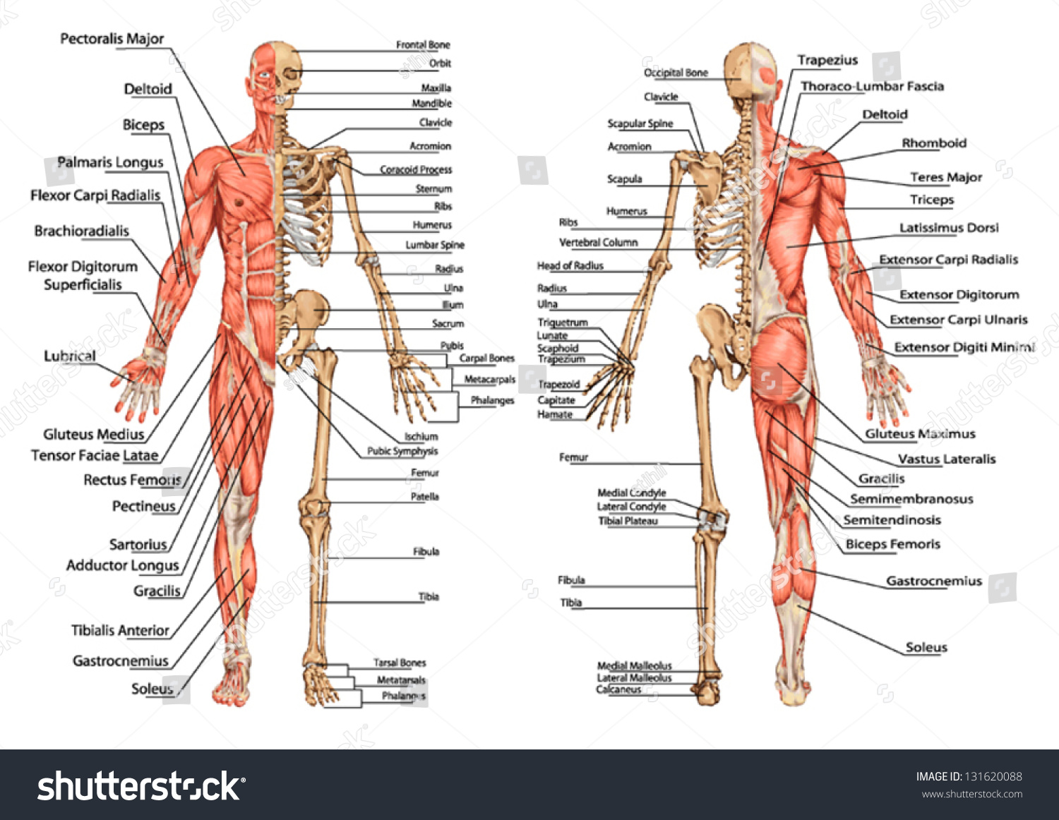
Skeletal System Carlson Stock Art Human skeletal system, Skeletal
The skull is a collection of 22 to 33 bones that protect the brain and many important structures of the head. Learn the complex anatomy of the skull by watch.
.PNG)
Skeletal System Presentation Biology
anterior view See anterior view in : french | spanish frontal bone lateral view of skull zygomatic bone Bone forming the cheek pouch and the outer edge of the eye socket. clavicle Long inward-curving bone located between the acromion and the sternum. scapula
human skeleton anterior view 744×1436 Anatomy System Human Body
fibula Long bone forming the outer portion of the leg located between the femur and the tarsus (foot bone). leg temporal bone Flat skull bone that protects mainly the organs responsible for hearing and equilibrium. lateral view of skull maxilla Toothed bone forming the upper jaw; it helps to form the palate, eye sockets and nasal fossae.
Human skeleton anterior view. Chromolithograph (?). Collection
Britannica Quiz The Human Body The central nervous system lies largely within the axial skeleton, the brain being well protected by the cranium and the spinal cord by the vertebral column, by means of the bony neural arches (the arches of bone that encircle the spinal cord) and the intervening ligaments.
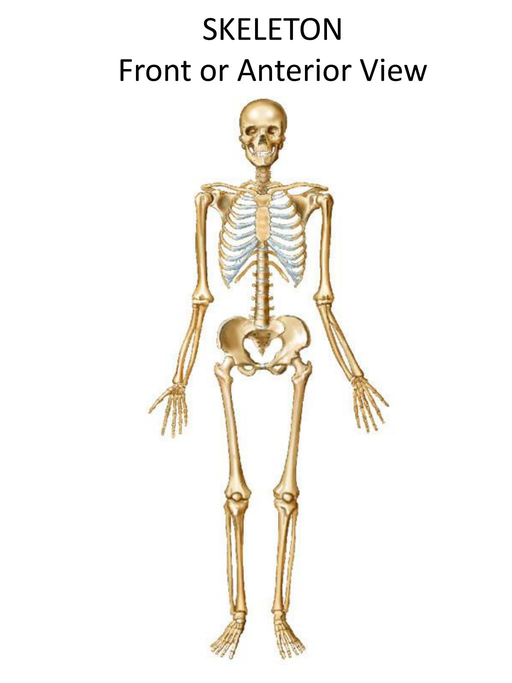
PPT SKELETON Front or Anterior View PowerPoint Presentation, free
The primary functions of the skeleton are to provide a rigid, internal structure that can support the weight of the body against the force of gravity, and to provide a structure upon which muscles can act to produce movements of the body. The lower portion of the skeleton is specialized for stability during walking or running.
LEE DOES ART. May 2013
The Skeletal System Explore the skeletal system with our interactive 3D anatomy models. Learn about the bones, joints, and skeletal anatomy of the human body. By: Tim Taylor Last Updated: Jul 29, 2020 2D Interactive NEW 3D Rotate and Zoom Anatomy Explorer HEAD AND NECK CHEST AND UPPER BACK PELVIS AND LOWER BACK ARM AND HAND LEG AND FOOT

34 Human Skeleton With Label Labels For Your Ideas
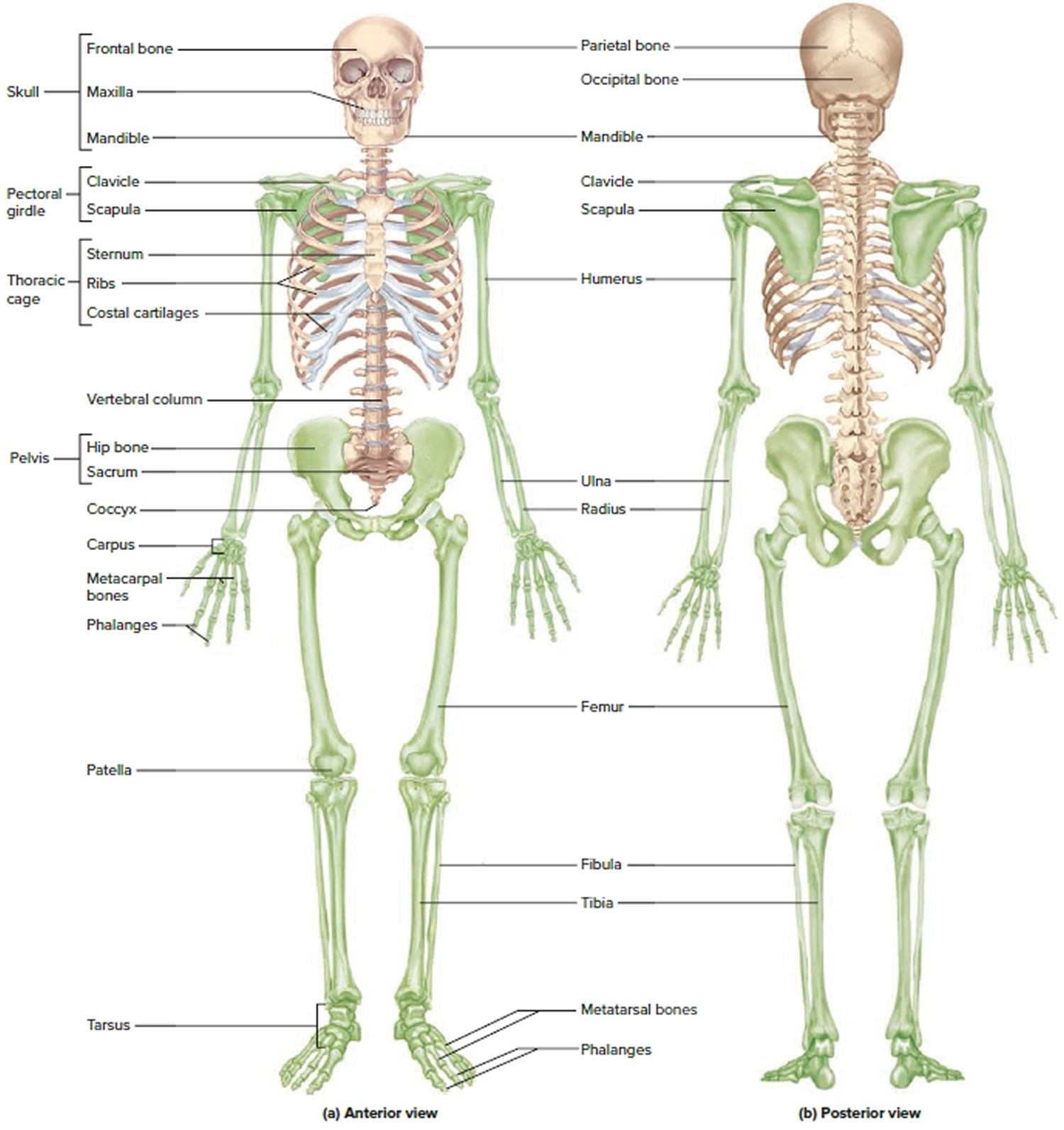
Identify three planes most commonly used in the study of anatomy; Distinguish between the posterior (dorsal) and the anterior (ventral) body cavities, identifying their subdivisions and representative organs found in each. Figure 1. Regions of the Human Body. The human body is shown in anatomical position in an (a) anterior view and a (b.
11. THE SKELETAL SYSTEM
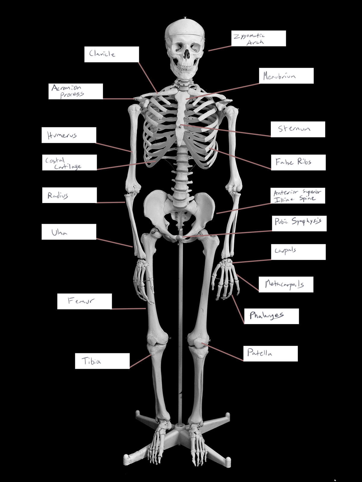
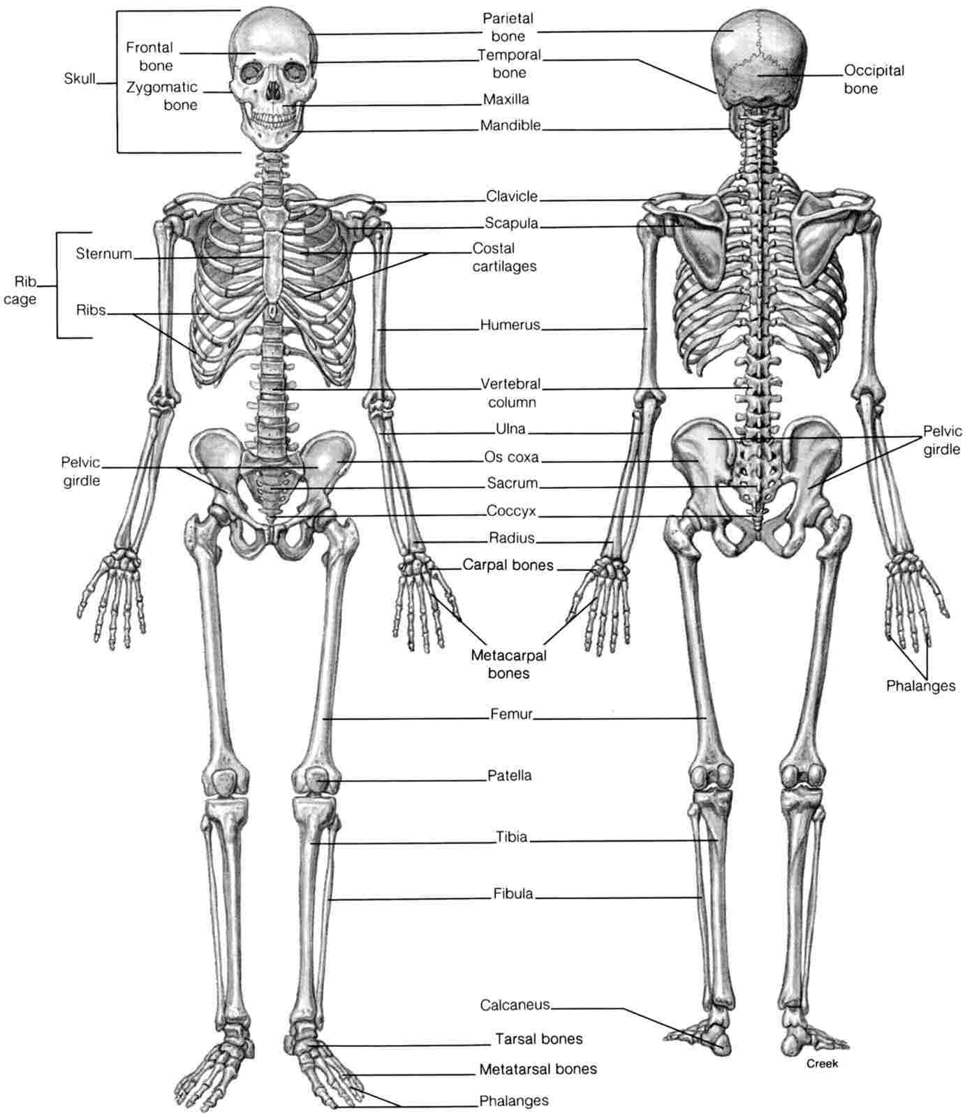
Anterior skeletal anatomy Overview The skeleton is made up of 206 bones in the adult and contributes to the form and shape of the body. The skeleton has several important functions for the body. The bones of the skeleton provide support for the soft tissues. For example, the rib cage supports the thoracic wall.
CrossFit The Skeleton Anterior and Posterior Views
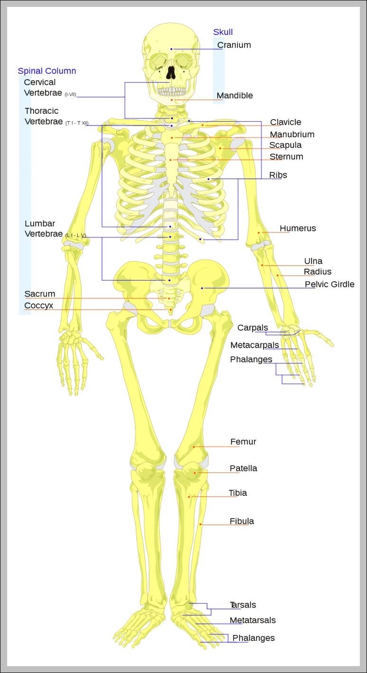
The vertebral column (spine or backbone) is a curved structure composed of bony vertebrae that are interconnected by cartilaginous intervertebral discs. It is part of the axial skeleton and extends from the base of the skull to the tip of the coccyx. The spinal cord runs through its center.

Human Skeleton Anterior View Images and Photos finder
Skeletal system (anterior view) From the moment you open your in the morning until you go to bed, you move around. Even during sleep you may fret, twist, and turn. Every movement that you do, no matter how inconspicuous, consists of a complex series of events with an equally complex
Human Skeletal System Drawing at Explore
RM K2298F - The human skeleton is made up of 206 articulated bones of varying sizes and shapes. RM 2DHWE55 - Human Skeleton, Anterior View (Finished Study for Table I), George Stubbs, 1724-1806, British, 1795 to 1806, Graphite on moderately thick, slightly textured, cream wove paper, Sheet: 21 3/8 x 16 inches (54.3 x 40.6 cm), anatomical.
lateral human anatomy
Figure 7.3.2 - Anterior View of Skull: An anterior view of the skull shows the bones that form the forehead, orbits (eye sockets), nasal cavity, nasal septum, and upper and lower jaws. Inside the nasal area of the skull, the nasal cavity is divided into halves by the nasal septum.

Anterior View Skeletal Diagram Skeletal System Worksheet, Human
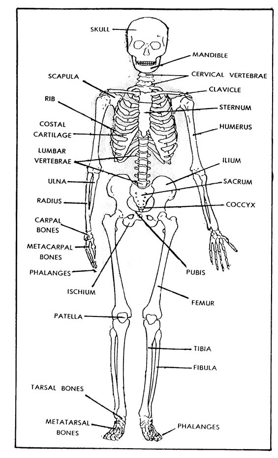
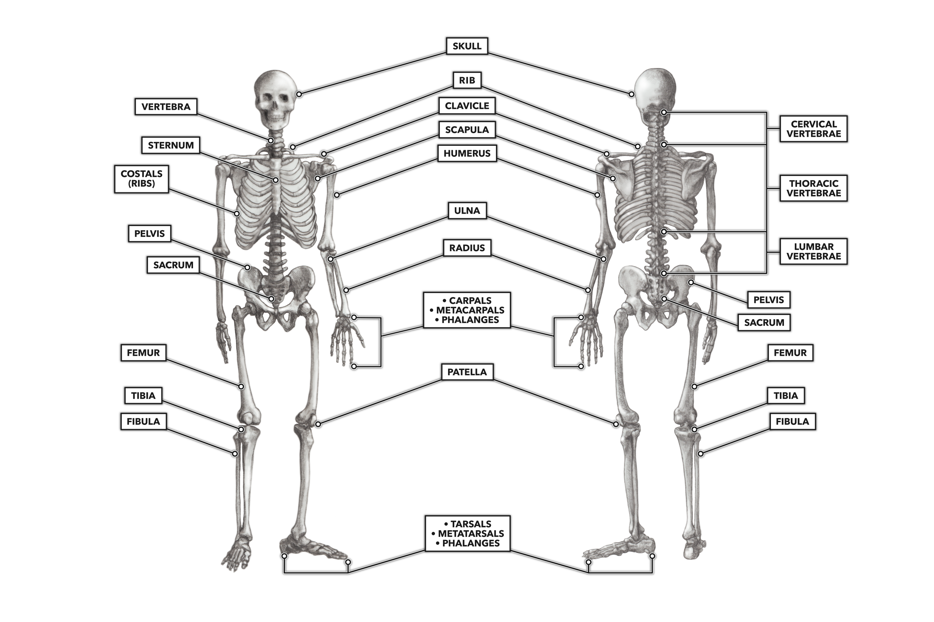
Anterior (left) and posterior (right) views of the human skeleton The skeleton is an aggregate of many connected bones. Bones are hard but alive, so they grow through childhood and adapt during adulthood. Our bones provide the hardware for human movement. They act as internal scaffolding, supporting everything visceral and muscular.

Anterior Skeleton (skeletal system) Page 4
Thoracic wall The first step in understanding thorax anatomy is to find out its boundaries. The thoracic, or chest wall, consists of a skeletal framework, fascia, muscles, and neurovasculature - all connected together to form a strong and protective yet flexible cage.. The thorax has two major openings: the superior thoracic aperture found superiorly and the inferior thoracic aperture.
Human Skeleton Skeletal System Function, Human Bones
Figure 1.4.1 - Regions of the Human Body: The human body is shown in anatomical position in an (a) anterior view and a (b) posterior view. The regions of the body are labeled in boldface. A body that is lying down is described as either prone or supine.
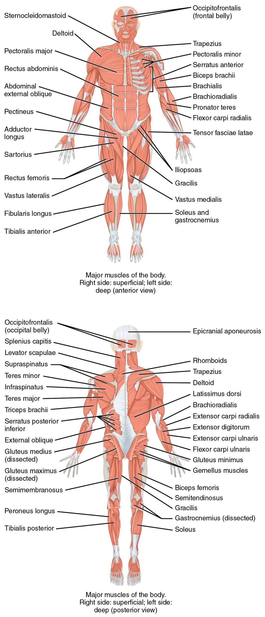
12.3 Types of Muscle Tissue Human Biology
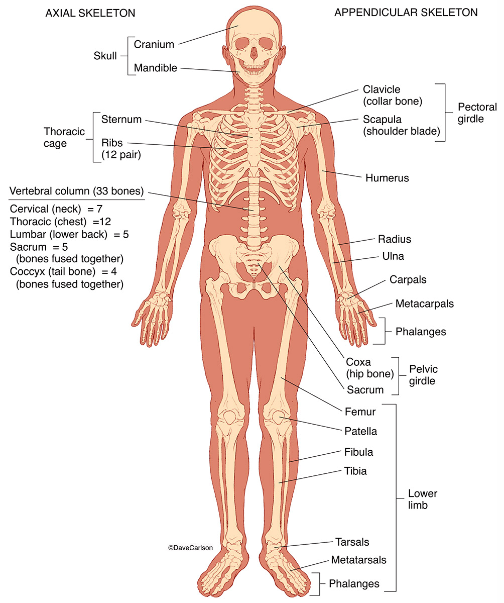
Skeleton & Spine. Shoulder & Back. Arm & Hand. Pelvis & Hip. Leg & Foot. Shaken Baby. The adult human skeleton is made up of 206 bones, and is divided into two main divisions: the axial and appendicular. Skeleton Axial and Appedicular. Skeleton & Spine.